San Luis Rey Equine Hospital offers a broad spectrum of state-of-the-art imaging technology in order to meet your horses specific needs and provide a rapid and accurate diagnosis. Techniques such as digital radiography, nuclear scintigraphy, computed tomography, endoscopy and ultrasound are commonly used to diagnose injury and illness in horses. Imaging is an exciting and rapidly changing field allowing an improved ability to assess the internal structures of the horse.
San Luis Rey Equine Hospital has a program in place for radiographs and nuclear scintigraphy results to be evaluated by a board certified radiologist.
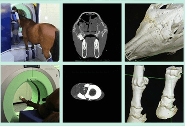
COMPUTED TOMOGRAPHY
S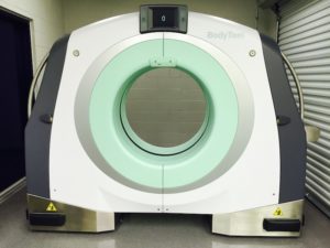 an Luis Rey Equine Hospital is proud to offer CT as part of our state-of-the-art diagnostic imaging services.
SLREH is one of only two clinics in North America to perform standing CT scans of the equine skull. This includes visualization of the brain, sinuses, nasal cavities, and teeth, all without the superimposition which greatly hampers standard radiography. With the installation design at SLREH, imaging of the skull is possible without the need for general anesthesia.
CT technology uses very small x-ray beams from many different angles around the body (called a slice) that are transmitted to a computer program, which produces an image of the highest quality. Images can be manipulated to render 3-dimensional reconstructions from 2-dimensional slices, producing not only exceptional images, but opening up whole new areas of diagnosis and treatment.
The CT unit in use at San Luis Rey Equine Hospital is able to accommodate very large body parts relative to the majority of CT units available for medical or veterinary use, measuring 85 cm in diameter relative to the much more common 60 cm diameter gantry. SLREH is the first veterinary hospital in the world to make use of this revolutionary, portable CT unit. The BodyTom from Samsung is currently used at a number of human hospitals in a variety of different applications including in the operating room, diagnostic units and emergency departments.
RADIOGRAPHY
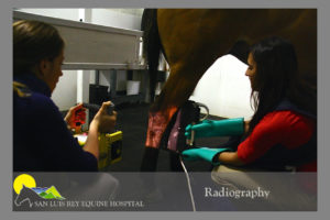 The hospital utilizes both computed and digital x-ray units, both which allow computerized enhancement of detail in specific regions of interest. Digital radiology units at the clinic provide high quality images of limbs and the cervical spine. We have a specially designed radiology room with a track in the ceiling allowing a large x-ray unit to be moved to almost any position around the horse, lessening the amount of stress to the patient. This large unit provides us with the ability to radiograph the abdomen, thorax, shoulder, lumbar spines, and hip that small radiograph machines are unable to penetrate.
Our radiograph viewing room boasts a large flat-screen TV to provide detailed image quality for client observation, with numerous anatomy specimens for teaching aids.
ABDOMINAL IMAGING
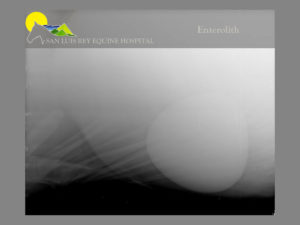
San Luis Rey Equine Hospital is equipped with a x-ray generator that can penetrate the large horse abdomen assisting in the diagnosis of enteroliths (stones) or sand colic. Our ultrasonography machines are also used for detailed imaging of the equine abdomen.
DIAGNOSTIC ULTRASOUND
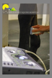
Ultrasonography is a specialized imaging technique used to examine soft tissues and bone surfaces. This procedure uses a hand held device, called a transducer, to send and receive high frequency sound waves. As the sound waves move into tissue, they are reflected back and transformed into an image that can be seen on a video screen. It is one of our most frequently used diagnostic tools and is used to diagnose conditions such as tendon, ligament and joint injuries, pneumonia, umbilical infections, colic and abdominal disease, cardiac disease, ocular issues and any swelling or mass, in addition to evaluating late term pregnancy. Ultrasound is commonly utilized at SLREH for guided biopsies of the kidney, liver, lung, or masses. At SLREH it is also utilized for ultrasound guided injections into bursas, tendons/ligaments, the sacroiliac space, and cervical facets. For advanced ultrasound imaging of complex cases or for echocardiography, SLREH offers expert consulting services from Dr. Tom Wilkinson at Washington State University.
NUCLEAR SCINTIGRAPHY
Commonly known as “bone scanning”, nuclear scintigraphy is an imaging technique allowing changes in metabolic activity of soft tissue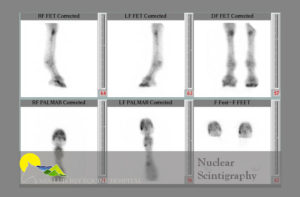 or bone to be visualized. It is one of the most commonly performed nuclear medicine procedures and is particularly useful in detecting bone inflammation in difficult to diagnose lameness cases. It is also valuable for patients with stress fractures, where a dynamic lameness exam is contraindicated. This procedure is highly sensitive for detecting early disease, allowing evaluation of the entire skeleton or a specific region, and the procedure is done without anesthesia.
A small amount of radioisotope pharmaceuticals are injected into the jugular vein through a catheter. These drugs become concentrated where bone is actively remodeling or undergoing change and identifies the suspected areas of injury. These “hot spots” are then imaged with radiographs and/or ultrasound for further evaluation.
We are proud to be one of the few private equine hospitals in the country to be able to offer this procedure, performing approximately 200 scans yearly.
MYELOGRAM
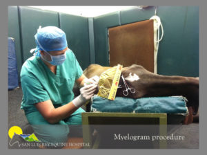
Myelograms are used to diagnose horses with cervical spinal cord compression or ‘Wobbler syndrome’. It is the most sensitive diagnostic tool utilized to diagnose horses with Wobbler Syndrome. The myelogram involves placing the horse under anesthesia, injecting a dye material into the vertebral column, and taking a series of radiographs with the horses neck in flexed and extended positions. The staff at SLREH have performed a large number of myelograms, and carry out this procedure in a quick and efficient manner, to reduce the amount of time for the horse to be under anesthesia.
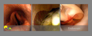
ENDOSCOPY
An endoscope is a small flexible fiber optic tube with a videocamera that allows direct visualization of internal structures. The endoscope allows examination and evaluation of otherwise inaccessible structures such as the upper respiratory tract (including the pharynx, trachea and guttural pouches), the upper gastrointestinal tract, the ears (otoscopy), the urethra and bladder as well as the cervix and uterus in the mare.
Videoendoscopy allows the image to be displayed on a monitor, in addition to providing video imaging capabilities for clients & referring veterinarians.